All referrals are accepted | Patient Portal
Call us now on 07 3209 5159
All referrals accepted | Patient Portal
Our Services

Queensland Radiology Specialists have a team of staff, dedicated to handling Worker’s Compensation cases. We ensure the process is efficient, providing a stress-free experience for both the referring doctor and the patient.

Nuclear medicine is a medical specialty that uses radioactive tracers to assess bodily functions and to diagnose and treat disease.

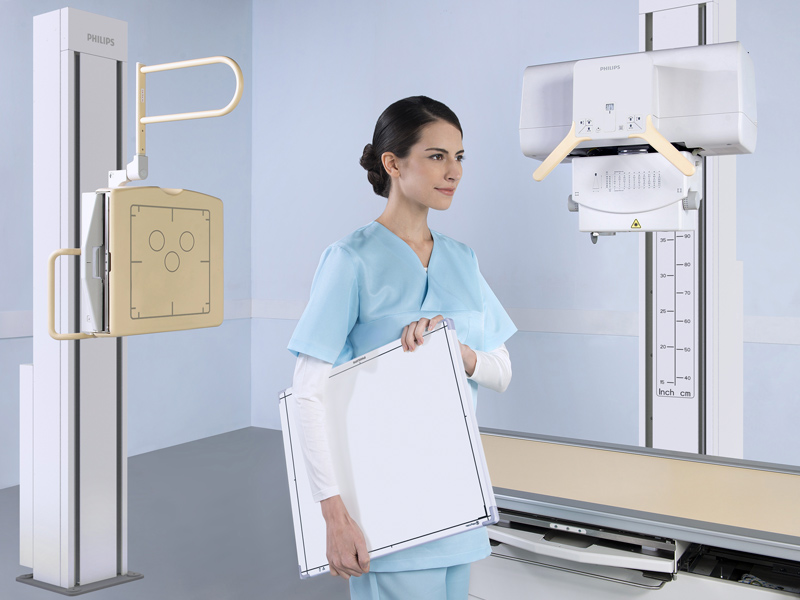
Radiography is an imaging technique using X-Rays, gamma rays, or similar radiation to view the internal form of an object.

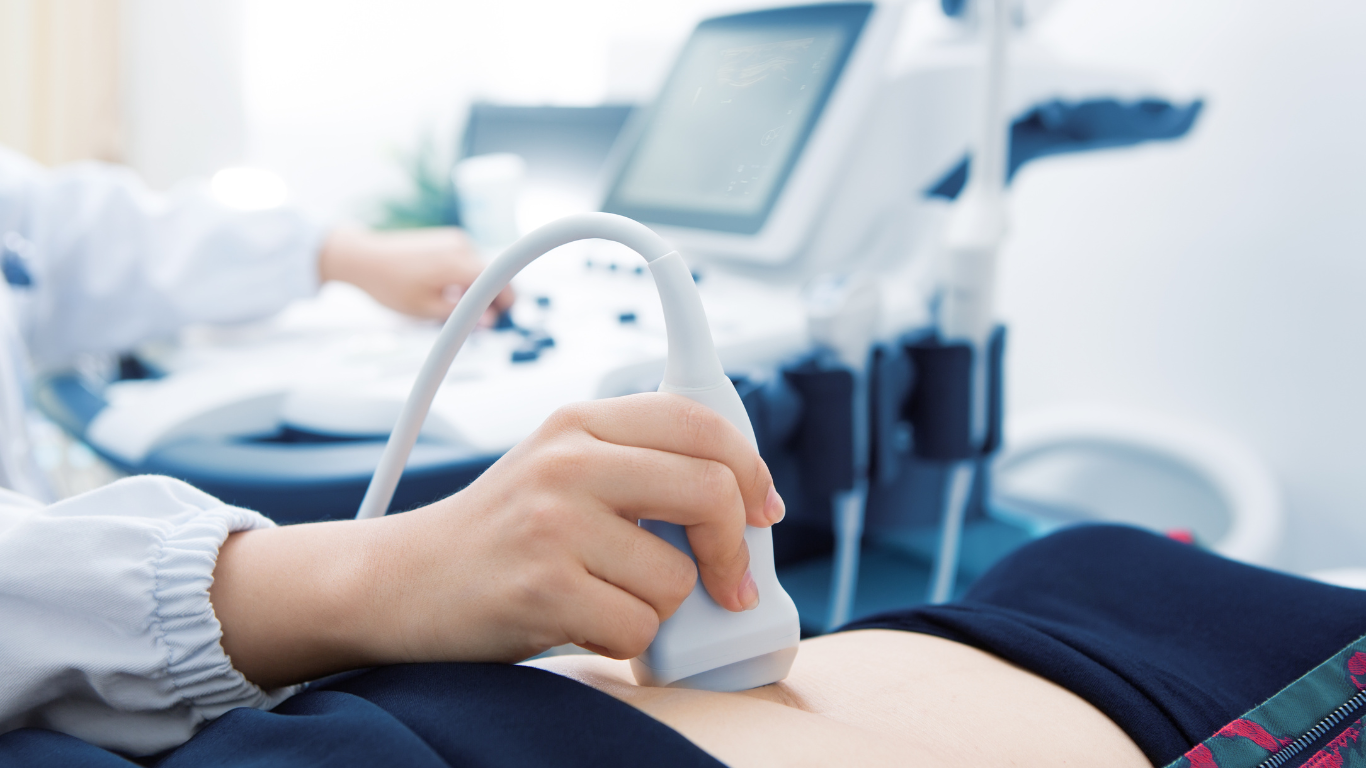
Ultrasound is used to create an image of internal body structures such as tendons, muscles, joints, blood vessels, and internal organs.

FNA and Core Biopsy – A Fine Needle Aspiration (FNA) is an interventional procedure used to acquire a cell sample for pathologic analysis. And a core biopsy involves the use of a specified instrument called a ‘core needle’, which is used to acquire tissue samples from a lesion.

MRI scanners use strong magnetic fields, magnetic field gradients, and radio waves to generate images of the organs in the body.

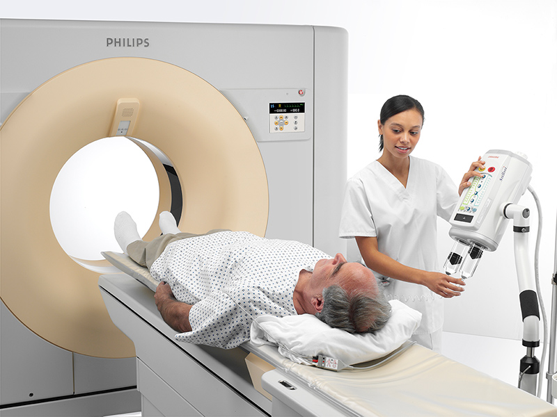
A CT scan makes use of combinations of X-ray measurements taken from different angles to produce cross-sectional images.

CTCA uses computed tomography (CT) scanning to detect blockages or narrowing of the coronary arteries.

An OPG is a panoramic X-ray of the upper and lower jaws, including the teeth. An OPG will take approximately 20 seconds.

Bone Mineral Densitometry uses a device to scan the lower spine and one of the hips to assess for possible osteoporosis or reduced bone density.

Body composition scans use DEXA technology to assess body fat, lean muscle and bone mass throughout the body.

In combination with your referring physician or pain management specialist, we offer a range of steroid and local anaesthetic injections.

In combination with your referring physician or pain management specialist, we offer a range of nerve blocks.

Platelet-rich plasma (PRP) therapy uses injections of a concentration of a patient’s own platelets to accelerate the healing of injured tendons and muscles.

Studies offered include dating , nuchal translucency and morphology ultrasounds.

Services are provided by CardioNexus.

Services are provided by CardioNexus.

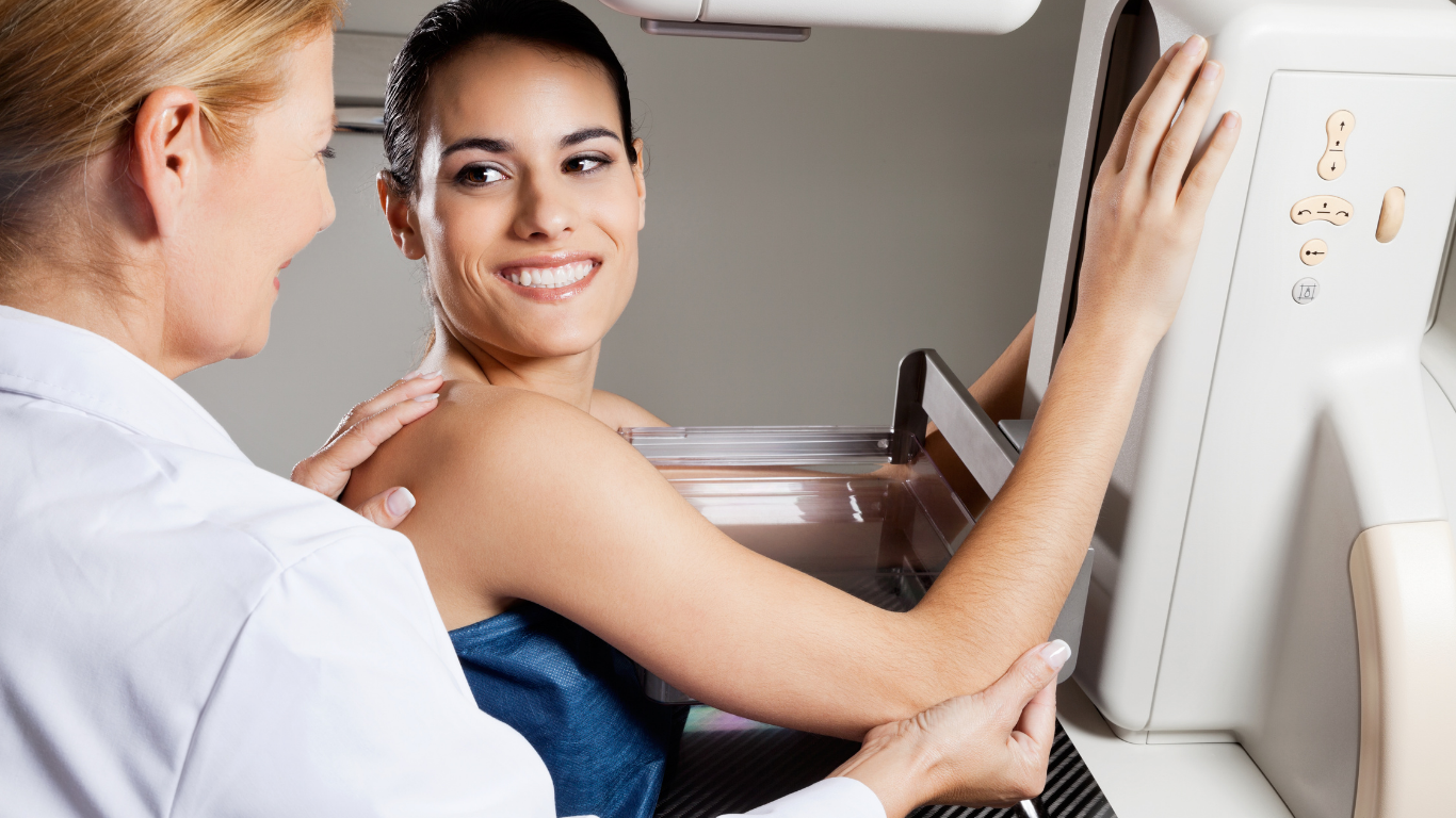
The goal of mammography is the early detection of breast cancer, typically through detection of characteristic masses or microcalcifications.

At Queensland Radiology Specialists, we understand the frustration that occurs when trying to arrange timely diagnostic imaging and interventional procedures for your patients on Worker’s Compensation.

Fortunately, we have a team of staff dedicated to handling Worker’s Compensation cases. We ensure the process is efficient, providing a stress-free experience for both the referring doctor and the patient. We will provide a prompt and accurate diagnosis so that the patient can start on the pathway to treatment, and ultimately, recovery.

Simply provide us a referral with the patient’s contact details, case number (if available) and:

      • We will contact the patient and prioritise their imaging / intervention
      • We accept all referrals
      • We will chase up approvals from insurers
      • All reports fast-tracked
      • We will treat you with the highest level of professional service and care


Please contact our WorkCover Team on:

Email: workcover@q-rad.com.au
Direct Line: 0455 191 067

Nuclear medicine is a medical specialty that uses radioactive tracers to assess bodily functions and to diagnose and treat disease. Specially designed cameras allow doctors to track the path of these radioactive tracers. Single Photon Emission Computed Tomography or SPECT is one of the most common imaging modalities in nuclear medicine.

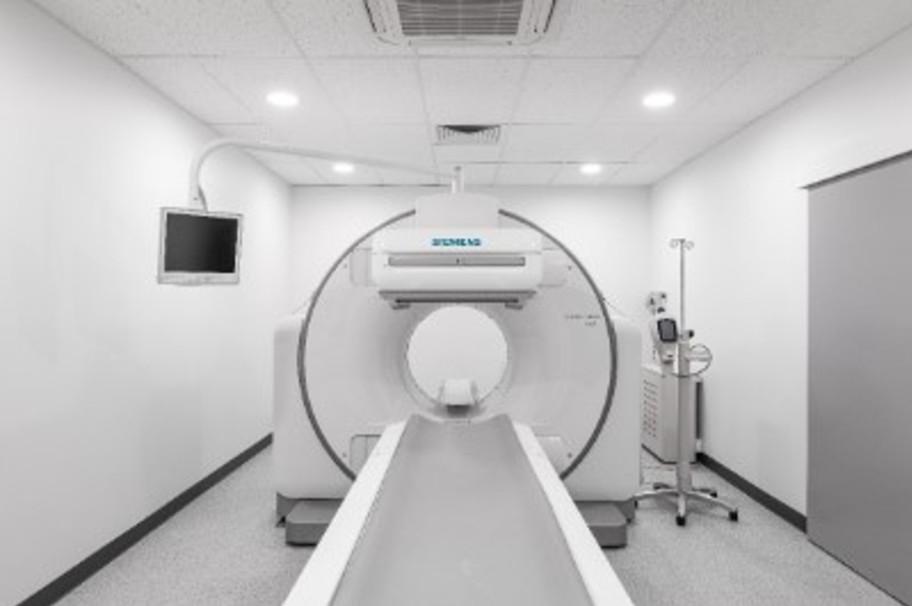
Our Symbia Intevo Bold combines high performance CT with advanced SPECT imaging to accommodate even the most challenging clinical questions.


At Queensland Radiology Specialists, we offer a bulk billed Nuclear Medicine service at our Mount Gravatt practice.

Bone Scan with SPECT CT:

      • Arthritis
      • Sport injuries
      • Suspected fractures with normal X-Ray
      • Identifying spinal facet joints suitable for injections


Myocardial Perfusion Scan (MPS)

      • Diagnosis of coronary artery disease and risk stratification
      • Preoperative assessment of patients at risk of ischemia and myocardial infarction
      • Assess lesions detected on CT coronary angiography for significance


Cerebral Perfusion Scan

      • Dementia


Thyroid Scan 

      • Hyperthyroidism (e.g. graves disease, thyroiditis, toxic adenomas)


Gall Bladder / Biliary Scan 

      • Acute and chronic cholecystitis
      • Post-cholecystectomy


Gastric Emptying Scan 

      • Investigate epigastric discomfort and bloating
      • Diagnosis and follow up of gastroparesis


Oesaophageal Transit and Reflux Scan 

      • Dysmotility
      • Reflex / aspiration


Renal Scans 

      • Assess and quantitate renal perfusion, differential renal function and drainage
      • Assess of presence of scaring


Qld Radiology Specialists is committed to providing ongoing essential services to all our patients and providers.

We are following all guidelines issued by the Department of Health to ensure the health and safety of our patients and employees.

We can all help in the process to protect against infection and prevent the virus spreading.


To ensure the safety to all, if you have travelled overseas in the last month or have a fever, cough, sore throat or shortness of breath, please call our practice on 07 3209 5159 prior to visiting.

X-ray services

Radiography is an imaging technique using X-Rays, gamma rays, or similar radiation to view the internal form of an object. To create the image, a beam of X-Rays or other form of electromagnetic radiation is produced by an X-Ray generator and is projected toward the object. A certain amount of the X-Rays or other radiation is absorbed by the object, dependent on the object’s density and structural composition. The X-Rays that pass through the object are captured behind the object by a detector (either photographic film or a digital detector). The generation of flat two dimensional images by this technique is called projectional radiography.

In computed tomography (CT scanning) an X-ray source and its associated detectors rotate around the subject which itself moves through the conical X-ray beam produced. Any given point within the subject is crossed from many directions by many different beams at different times. Information regarding attenuation of these beams is collated and subjected to computation to generate two dimensional images in three planes (axial, coronal, and sagittal), which can be further processed to produce a three dimensional image.

X-Ray services for Southeast Queensland

At Queensland Radiology Specialists, you do not require an appointment for an X-Ray. Walk-ins are welcome at all local practices.

Ultrasound services

Medical ultrasound (also known as diagnostic sonography or ultrasonography) is a diagnostic imaging technique based on the application of ultrasound. It is used to create an image of internal body structures such as tendons, muscles, joints, blood vessels, and internal organs. Its aim is often to find a source of a disease or to exclude pathology. The practice of examining pregnant women using ultrasound is called obstetric ultrasound, and was an early development and application of clinical ultrasonography.

How Do Ultrasounds Work?

Ultrasound refers to sound waves with frequencies which are higher than those audible to humans (>20,000 Hz). Ultrasonic images, also known as sonograms, are made by sending pulses of ultrasound into tissue using a probe. The ultrasound pulses echo off tissues with different reflection properties and are recorded and displayed as an image.

Many different types of images can be formed. The commonest is a B-mode image (Brightness), which displays the acoustic impedance of a two-dimensional cross-section of tissue. Other types can display blood flow, motion of tissue over time, the location of blood, the presence of specific molecules, the stiffness of tissue, or the anatomy of a three-dimensional region.

Compared to other dominant methods of medical imaging, ultrasound has several advantages. It provides images in real-time and is portable and can be brought to the bedside. It is substantially lower in cost than other imaging modalities and does not use harmful ionizing radiation. Drawbacks include various limits on its field of view, such as the need for patient cooperation, dependence on physique, difficulty imaging structures behind bone and air, and the necessity of skilled operators at Queensland Radiology Specialists.

Ultrasound services for Southeast Queensland

At Queensland Radiology Specialists, we are continuing to offer a range of bulk billing ultrasound services for Medicare eligible patients.

Secure your same day appointment with Queensland Radiology Specialists by calling 1300 201 888 or filling out an enquiry form.

Fine Needle Aspiration

A Fine Needle Aspiration (FNA) is an interventional procedure used to acquire a cell sample for pathologic analysis. The specimen is extracted (aspirated) by the insertion of a fine needle (typically smaller than that used to take blood), in the region of interest with cells being acquired through the gentle manipulation of the needle. To ensure accuracy of the sample taken, ultrasound is used by the radiologist to correctly locate and place the needle in the lesion whilst the sample is acquired. Local anesthetic will be first be administered to the region of interest site prior to the acquisition of the first biopsy. For proper analysis to be made by the pathologist once back at the laboratory, multiple samples or ‘passes’ are taken to ensure enough cells are acquired. An on-site cytologist will be present to assess each acquired sample under a microscope, to reduce the likelihood of inconclusive results due to insufficient cell number. Depending on the pathology provider used, result time may vary, however typically they will be available to you referring doctor within 2-3 business days.

What can I expect following the procedure?

Any pain after FNA can be relieved with non-prescription analgesics, such as paracetamol. You should not take aspirin, because this thins the blood and may increase the risk of bleeding and bruising. Minimal bleeding or bruising can appear after the procedure, especially if you are taking anticoagulant medication, aspirin or fish oil. This can be minimised by good compression and the application of an icepack on the skin at the site of the FNA. Breast FNA does not leave a scar on the skin and there is a low risk of infection.

Core Biopsy / Vacuum Assisted Core Biopsies

A core biopsy involves the use of a specified instrument called a ‘core needle’, which is used to acquire tissue samples from a lesion. Following the administration of local anesthetic to the skin a small incision is made (typically less than 5mm) which allows the core needle to be easily advanced through the skin surface to the lesion in question. Once appropriately positioned the core needle is fired through the lesion making an audible ‘click’ as the sample is acquired. Depending on the efficacy of the sample, the process may be repeated multiple times to ensure that suitable samples are obtained for assessment back at the laboratory.
Due to the small incision required in the skin, a fine scar may remain (typically only a few mm) which is likely to fade in time.

What can I expect following the procedure?

Following the core biopsy, you may experience some localised bruising and discomfort which may last for several days. If you require symptomatic relief from any pain experienced you can take non- prescription analgesics, such as paracetamol. To avoid exacerbation of bruising, avoid vigorous exercise for 24 hours post procedure and the use of Aspirin for pain relief due to its blood thinning properties. Ice packs and compression may also be utilised at the biopsy site to relive pain and mitigate any additional bruising.

MRI is a medical imaging technique used in radiology to form pictures of the anatomy and the physiological processes of the body in both health and disease. MRI scanners use strong magnetic fields, magnetic field gradients, and radio waves to generate images of the organs in the body.

MRI was originally called NMRI (nuclear magnetic resonance imaging), but the use of ‘nuclear’ in the acronym was dropped to avoid negative associations with the word. Certain atomic nuclei are able to absorb and emit radio frequency energy when placed in an external magnetic field. In clinical and research MRI, hydrogen atoms are most often used to generate a detectable radio-frequency signal that is received by antennas in close proximity to the anatomy being examined. Hydrogen atoms are naturally abundant in people and other biological organisms, particularly in water and fat. For this reason, most MRI scans essentially map the location of water and fat in the body. Pulses of radio waves excite the nuclear spin energy transition, and magnetic field gradients localize the signal in space. By varying the parameters of the pulse sequence, different contrasts may be generated between tissues based on the relaxation properties of the hydrogen atoms therein.

While the hazards of X-Rays are now well-controlled in most medical contexts, an MRI scan may still be seen as a better choice than a CT scan. MRI is widely used in hospitals and clinics for medical diagnosis, staging of disease and follow-up without exposing the body to radiation. An MRI may yield different information compared with CT. There may be risks and discomfort associated with MRI scans. Compared with CT scans, MRI scans typically take longer and are louder, and they usually need the subject to enter a narrow, confining tube.

MRI services for Southeast Queensland

At Queensland Radiology Specialists, we are thrilled to share our most advanced MRI machine to date, the Siemens MAGNETOM Altea.

Our MAGNETOM Altea, with state of the art A.I Deep Resolve Technology, ensures best patient experience and provides superior image quality to increase diagnostic accuracy.

      • High quality personalised scans
      • Cutting edge acceleration technologies to deliver up to 50% faster examination times (10-15 minutes for routine clinical examinations)
      • Effective noise reduction and scan progressed delivered throughout
      • Wide bore technology to help reduce claustrophobia

Queensland Radiology Specialists offer bulk billing MRI services for DVA Gold Cards and WorkCover patients on approval.

Secure your same day appointment with Queensland Radiology Specialists by calling 1300 201 888 or filling out an enquiry form.

CT Scan services

A Computed Tomography (CT) and formerly known as a computerized axial tomography scan or CAT scan, makes use of computer-processed combinations of many X-ray measurements taken from different angles to produce cross-sectional (tomographic) images (virtual slices) of specific areas of a scanned object, allowing the user to see inside the object without cutting.

Digital geometry processing is used to further generate a three-dimensional volume of the inside of the object from a large series of two-dimensional radiographic images taken around a single axis of rotation. Medical imaging is the most common application of X-Ray CT. Its cross-sectional images are used for diagnostic and therapeutic purposes in various medical disciplines.

The CT scan is often used to refer to X-Ray CT, because it is the most commonly known form. But, many other types of CT exist, such as positron emission tomography (PET) and single-photon emission computed tomography (SPECT). X-Ray tomography, a predecessor of CT, is one form of radiography, along with many other forms of tomographic and non-tomographic radiography.

CT produces data that can be manipulated in order to demonstrate various bodily structures based on their ability to absorb the X-ray beam. Although, historically, the images generated were in the axial or transverse plane, perpendicular to the long axis of the body, modern scanners allow this volume of data to be reformatted in various planes or even as volumetric (3D) representations of structures.

CT Colonoscopy

Virtual colonoscopy (VC), also called CT colonography or CT pneumocolon, is a medical imaging procedure which uses X-Rays and computers to produce two and three-dimensional images of the colon (large intestine) from the lowest part, the rectum, all the way to the lower end of the small intestine and display them on a screen. The procedure is used to diagnose colon and bowel disease, including polyps, diverticulosis and cancer. VC is performed via CT. It may be performed following a conventional or optic colonoscopy (OC). Both OC and VC require bowel preparation with laxatives. The bowel requires insufflation with air using a tube placed in the rectum before the procedure. A muscle relaxant is given and there may be some discomfort to inflate the nose enough to allow optimal viewing. A virtual colonoscopy can provide 3D reconstructed endoluminal views of the bowel.

CT Scan services for Southeast Queensland

At Queensland Radiology Specialists, we are continuing to offer a range of bulk billing CT Scan services for Medicare eligible patients.

Secure your same day appointment with Queensland Radiology Specialists by calling 1300 201 888 or filling out an enquiry form.

What is CTCA?

CTCA uses computed tomography (CT) scanning to image of the coronary arteries and the heart. These arteries supply blood to the heart muscle and disease of these arteries, atherosclerosis is responsible for most heart attacks. CTCA detects blockages or narrowing of the coronary arteries.

What is the radiation dose?

Radiation dosage vary between different CT scanners. The benefits of CTCA at Queensland Radiology Specialists is that we uses the Philips Elite 256-slice CT scanner which allows the scan to be performed with high quality and at the lowest possible radiation dose.

How long does a CTCA take?

Most CTCA takes between 30 minutes and 2 hours. The CTCA test is carried out in three parts which includes preparation, scanning and recovery. Timing varies and although the scan usually take approximately 20-30 minutes, the work up process is quite involved. It is recommended that you allow a couple of hours for the examination in case you require medication to slow your heart rate for the scan.

How do I prepare for the scan?

Your doctor may give you a drug to slow down your heart rate before coming for your scan. When making your appointment, we will ask you to:

      • Avoid caffeine (tea, coffee, energy drinks,carbonated soft drinks) morning prior to the scan
      • Do not take any medication for erectile dysfunction for 72 hours prior to your CT, including viagra
      • Fast (nothing to eat or drink) for 2 hours prior to the scan


Please tell booking staff if:

      • You are pregnant (or think you may be pregnant)
      • If you have any allergies (especially to iodine)


What do I need to bring?

      • Medicare card
      • Request form
      • Any previous imaging scans that you make have had


On the day of your appointment, it is important to arrive 60 minutes prior to your examination for a pre-exam assessment which includes monitoring of blood pressure and heart rate to determine if you require any medication to slow down the heart beat (beta blocker) before your scan. If beta-blocker is required, it will take approximately 1 hour for the medication to take effect. Your CTCA examination will be performed once the doctor or CT radiographer is satisfied with your heart rate.

What can I expect during the scan?

You will have an intravenous cannula inserted into one of your arm veins and ECG leads placed on your chest. You will then be taken into the CT scanner room.

You will lie on a bed and nitroglycerin (GTN) will be sprayed under your tongue in order to expand and relax your coronary arteries. Occasionally GTN can cause a transient headache. While you are on the bed, you will be given a rapid IV injection of iodine contrast agent through the cannula. You may notice a warm sensation and a metallic taste in your mouth during the contrast injection which is a normal sensation. When the iodine contrast reaches the heart through the veins, the scan is started and images of the arteries and heart are acquired.

It is important to lie very still during the scan. If you move the images will be blurred and we may not be able to use them and scan may be repeated.

What happens following the scan?

Your blood pressure and heart rate will be checked after the scan. If your blood pressure and heart rate are normal and you feel well, we will take the cannula out of your arm and you will be allowed to leave.

In some cases, we may keep you for a while to give you something to eat and drink and/or to give you fluids through your veins (if you are dehydrated).

If you feel unwell, please tell a staff member as soon as possible.

When will I get my results?

A radiologist will write a report. The scan results and report will be sent to your referring doctor. Please contact your doctor to discuss or obtain a copy of the results. Your results will not be available at the end of your scan.

X-Rays use radiation to take pictures of bones and other parts inside the body. An (Orthopantomogram) OPG is a panoramic X-Ray of the upper and lower jaws, including the teeth. The OPG unit is specifically designed to rotate around the patient’s head during the scan. An OPG will take approximately 20 seconds.

An OPG can be used to look for:

      • Fractures
      • Dislocated jaw
      • Infection
      • Dentition (teeth)
      • It can also be used for surgical planning


Your doctor, dentist or dental specialist knows the risks of having an OPG and will consider the risks before recommending you have this type of X-Ray.

Bring your referral letter or request form and all OPG X-Rays taken within the last 2 years with you. Leave the X-Rays with the medical imaging staff as the doctor may need to look at them. The staff member will tell you when these are ready to be picked up.

Medical imaging staff will ask you to sit on a chair or stand for the OPG. It is important that you tell the staff if you have difficulty sitting or standing unassisted. The radiographer may place a protective shield over the parts of your body not being X-Rayed, or you may be asked to wear a protective apron.

When you are ready, the radiographer will go behind a screen to start the OPG machine. They will ask you to be still during the OPG. When your OPG is finished you will be asked to wait while the radiographer checks the pictures. The procedure usually takes about 5 minutes including time taken to get ready.


Dental X-Ray services for Southeast Queensland

At Queensland Radiology Specialists, we are continuing to offer a range of bulk billing dental services for Medicare eligible patients.

Secure your same day appointment with Queensland Radiology Specialists by calling 1300 201 888 or filling out an enquiry form.

START TYPING AND PRESS ENTER TO SEARCH