All referrals are accepted | Patient Portal
Call us now on 07 3209 5159
All referrals accepted | Patient Portal
Our Services

Queensland Radiology Specialists have a team of staff, dedicated to handling Worker’s Compensation cases. We ensure the process is efficient, providing a stress-free experience for both the referring doctor and the patient.

Nuclear medicine is a medical specialty that uses radioactive tracers to assess bodily functions and to diagnose and treat disease.

Radiography is an imaging technique using X-Rays, gamma rays, or similar radiation to view the internal form of an object.

Ultrasound is used to create an image of internal body structures such as tendons, muscles, joints, blood vessels, and internal organs.

FNA and Core Biopsy – A Fine Needle Aspiration (FNA) is an interventional procedure used to acquire a cell sample for pathologic analysis. And a core biopsy involves the use of a specified instrument called a ‘core needle’, which is used to acquire tissue samples from a lesion.

MRI scanners use strong magnetic fields, magnetic field gradients, and radio waves to generate images of the organs in the body.

A CT scan makes use of combinations of X-ray measurements taken from different angles to produce cross-sectional images.

CTCA uses computed tomography (CT) scanning to detect blockages or narrowing of the coronary arteries.

An OPG is a panoramic X-ray of the upper and lower jaws, including the teeth. An OPG will take approximately 20 seconds.

Bone Mineral Densitometry uses a device to scan the lower spine and one of the hips to assess for possible osteoporosis or reduced bone density.

Body composition scans use DEXA technology to assess body fat, lean muscle and bone mass throughout the body.

In combination with your referring physician or pain management specialist, we offer a range of steroid and local anaesthetic injections.

In combination with your referring physician or pain management specialist, we offer a range of nerve blocks.

Platelet-rich plasma (PRP) therapy uses injections of a concentration of a patient’s own platelets to accelerate the healing of injured tendons and muscles.

Studies offered include dating , nuchal translucency and morphology ultrasounds.

Services are provided by CardioNexus.

Services are provided by CardioNexus.

The goal of mammography is the early detection of breast cancer, typically through detection of characteristic masses or microcalcifications.

Bone Mineral Densitometry, also commonly referred to as BMD, is the examination that uses a device to scan the lower spine and one of the hips (or wrist if a hip replacement has been performed), to assess for possible osteoporosis or reduced bone density. Osteoporosis results from loss of bone mass, leading to bone weakening and increasing the chance of a fracture. Results will be compared with previous studies and hence it is important to have these available. Ideally, your BMD should be performed on the same scanner each time to provide the most accurate assessment over time.

Bone mass increases from infancy until about 20-30 years when normal young adult achieves peak bone mass. After this time bone mass gradually decreases with age, and this loss of bone mass is further accelerated in females related to menopause. Once bone mass falls below a certain threshold, the risk of fracture dramatically increases.

In most cases we can fit you in on the day without an appointment. No specific preparation is necessary for this test. The test should not be performed within a period of 72 hours following a nuclear bone scan or within one week following a barium study (barium meal, small bowel barium follow-through or barium enema).

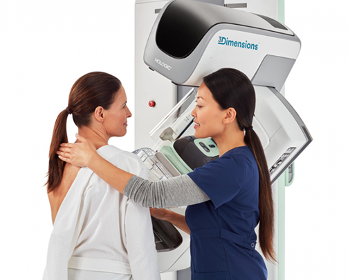
We may ask you to change into a gown to avoid clothing creating confusing shadows on the image. Clothing without metal (e.g. zips, buttons) should be worn for the examination.

You will be required to lie on your back on the scanner table. You will hear minimal noise when the scanner moves above you. There will be no injections or discomfort and no breath hold is required. However, it is important to remain as still as possible for the examination.

Following the scan, the information obtained is processed by computer and reported by the radiologist or nuclear medicine physician. The scan takes only a few minutes but the examination can take up 15 minutes to complete as we ask some questions about medical history.

The cost of the scan may be claimed from Medicare under certain circumstances. Unfortunately, these criteria are very strict and some patients are not eligible to claim a Medicare rebate and are therefore required to pay a fee at the time of consultation – this will be discussed at the time of booking.

Body composition scans use DEXA (Dual X-ray Energy Absorptiometry) technology in assessing body fat, lean muscle and bone mass throughout the body. No preparation is required and it is a good way to assess your progress with your fitness goals. The study should not be performed if you are pregnant or breast feeding.

Following the examination, you will receive a personalised breakdown regarding amount and distribution of fat, muscle, bone and water in the body.

In combination with your referring physician or pain management specialist, we offer a range of steroid and local anaesthetic injections including:

        • Subacromial subdeltoid bursal injections
        • Trochanteric bursal injections
        • Tendon sheath injections
        • Carpal tunnel injections
        • Sacroiliac joint injections
        • Facet joint injections
        • Costovertebral joint injection
        • Sternoclavicular joint injection
        • Large and small joint injections


What can I expect following the procedure?

As with any injection, there is sometimes a dull ache for a few hours after the procedure. There might be an area of numbness around the injection site, for 1 or 2 hours, due to the local anaesthetic. Some minor localised bruising may be noted, which will be minimalised by the application of light pressure and a dressing following the procedure.

The corticosteroid does not usually start working for 24 hours, and gradual improvement may be noted over the coming days to weeks. During this time, the normal symptoms might continue or occasionally worsen. If symptoms are much worse, it generally indicates a reaction to part of the injected medication or to the injection itself. If you find this worrying or distressing, you should see your own doctor or contact our practice.

Sometimes people can experience general reactions, such as flushing and redness of the body and face, related to the absorption of the corticosteroid into the body. These occur over the first few days.

Insulin dependent diabetic patients may notice a moderate rise in blood sugar for up to 10 days after injection. You may need to temporary adjust your medications as appropriate. Please consult your diabetic / management doctor if concerned.

In combination with your referring physician or pain management specialist, we offer a range of nerve blocks including:

      • Cervical nerve root blocks
      • Lumbar nerve root blocks
      • Occipital nerve blocks
      • Cluneal nerve blocks
      • Stellate ganglion nerve blocks

Platelet-rich plasma (PRP) therapy, uses injections of a concentration of a patient’s own platelets to accelerate the healing of injured tendons and muscles. In this way, PRP injections use each individual patient’s own healing system to improve musculoskeletal problems.

How long does it take to work?

PRP does not offer quick pain relief. Pain gradually subsides as the injured tissue repairs and pain relieving factors activate. This can take weeks or months, but when successful, the effect is lasting. If relief is not sufficient at 3 months, a second injection may be performed.

What is the process?

After a referral from your doctor, a consultation with the radiologist will take place along with physical examination and review of your imaging to determine if PRP injection is appropriate. Non steroidal anti-inflammatory medications (NSAID’s) including Nurofen, Voltaren, Mobic must be stopped 10 days prior to the procedure because these interfere with platelet production by the bone marrow

A quantity (up to 20mls) of autologous blood will be extracted by venesection, usually from an arm vein. The blood will be centrifuged for up to 15 minutes. The PRP is separated from the red blood cells and injected under US guidance to the area of injury.

What can I expect following the procedure?

Following the injection, you may experience some localised bruising and discomfort which can be relieved with compression ice packs and non-prescription analgesia. The effects of the injection may not be noted for 4-6 weeks following the procedure as the regenerative benefits from the platelets act upon the affected area.

Given the injection involves an autologous substance (derived from your own body) the risk of reaction and adverse effects are extremely minimal making the PRP injection a safe alternative to other pain management and reparative therapies.

Depending on the desired approach, a rehabilitation program may be provided to you by your referring doctor or physiotherapist which may be undertaken following the procedure. Follow up injections may also be performed if required, with the number and time frame being determined in consultation with your doctor.

Studies offered:

      • First trimester dating ultrasound
      • Nuchal translucency ultrasound
      • Third trimester morphology ultrasound

Services are provided by CardioNexus.

Services are provided by CardioNexus.

Mammography (also called Mastography) is the process of using low-energy X-Rays (usually around 30 kVp) to examine the human breast for diagnosis and screening. The goal of mammography is the early detection of breast cancer, typically through detection of characteristic masses or microcalcifications.

As with all X-Rays, mammograms use doses of ionizing radiation to create images. These images are then analyzed for abnormal findings. It is usual to employ lower-energy X-Rays, typically Mo (K-shell X-Ray energies of 17.5 and 19.6 keV) and Rh (20.2 and 22.7 keV) than those used for radiography of bones. Ultrasound, ductography, positron emission mammography (PEM), and magnetic resonance imaging (MRI) are adjuncts to mammography. Ultrasound is typically used for further evaluation of masses found on mammography or palpable masses not seen on mammograms. Ductograms are still used in some institutions for evaluation of bloody nipple discharge when the mammogram is non-diagnostic. MRI can be useful for further evaluation of questionable findings, as well as for screening pre-surgical evaluation in patients with known breast cancer, in order to detect additional lesions that might change the surgical approach, for example, from breast-conserving lumpectomy to mastectomy. Other procedures being investigated include tomosynthesis.

For the average woman, the U.S. Preventive Services Task Force recommends (2016) mammography every two years between the ages of 50 and 74, concluding that “the benefit of screening mammography outweighs the harms by at least a moderate amount from age 50 to 74 years and is greatest for women in their 60s”. The American College of Radiology and American Cancer Society recommend yearly screening mammography starting at age 40. The Canadian Task Force on Preventive Health Care (2012) and the European Cancer Observatory (2011) recommend mammography every 2 to 3 years between ages 50 and 69. These task force reports point out that in addition to unnecessary surgery and anxiety, the risks of more frequent mammograms include a small but significant increase in breast cancer induced by radiation. Additionally, mammograms should not be performed with increased frequency in patients undergoing breast surgery, including breast enlargement, mastopexy, and breast reduction.

What is Mammography?

Diagnostic Mammography is a specialised type of low dose X-Ray of the breasts used to help diagnose breast cancer in women who have symptoms such as pain or a lump in their breast. Diagnostic mammography may also be done after an abnormal screening mammogram in order to evaluate an area of concern.

How is 3D Mammography different?

3D mammography is an advanced form of breast imaging where multiple images of the breast from different angles are captured and reconstructed into a three-dimensional image set. The 3D mammogram machine creates both 3D images and standard 2D mammogram images because both types of images have some advantages in seeing certain breast abnormalities.

3D mammograms can:

      • Reduce the need for follow-up imaging
      • Improve breast cancer detection, especially in dense breast tissue
      • Reduce the number of unnecessary biopsies

START TYPING AND PRESS ENTER TO SEARCH