
Mammography
Mammography (also called Mastography) is the process of using low-energy X-Rays (usually around 30 kVp) to examine the human breast for diagnosis and screening. The goal of mammography is the early detection of breast cancer, typically through detection of characteristic masses or microcalcifications.
As with all X-Rays, mammograms use doses of ionizing radiation to create images. These images are then analyzed for abnormal findings. It is usual to employ lower-energy X-Rays, typically Mo (K-shell X-Ray energies of 17.5 and 19.6 keV) and Rh (20.2 and 22.7 keV) than those used for radiography of bones. Ultrasound, ductography, positron emission mammography (PEM), and magnetic resonance imaging (MRI) are adjuncts to mammography. Ultrasound is typically used for further evaluation of masses found on mammography or palpable masses not seen on mammograms. Ductograms are still used in some institutions for evaluation of bloody nipple discharge when the mammogram is non-diagnostic. MRI can be useful for further evaluation of questionable findings, as well as for screening pre-surgical evaluation in patients with known breast cancer, in order to detect additional lesions that might change the surgical approach, for example, from breast-conserving lumpectomy to mastectomy. Other procedures being investigated include tomosynthesis.
For the average woman, the U.S. Preventive Services Task Force recommends (2016) mammography every two years between the ages of 50 and 74, concluding that “the benefit of screening mammography outweighs the harms by at least a moderate amount from age 50 to 74 years and is greatest for women in their 60s”. The American College of Radiology and American Cancer Society recommend yearly screening mammography starting at age 40. The Canadian Task Force on Preventive Health Care (2012) and the European Cancer Observatory (2011) recommend mammography every 2 to 3 years between ages 50 and 69. These task force reports point out that in addition to unnecessary surgery and anxiety, the risks of more frequent mammograms include a small but significant increase in breast cancer induced by radiation. Additionally, mammograms should not be performed with increased frequency in patients undergoing breast surgery, including breast enlargement, mastopexy, and breast reduction.
What is Mammography?
Diagnostic Mammography is a specialised type of low dose X-Ray of the breasts used to help diagnose breast cancer in women who have symptoms such as pain or a lump in their breast. Diagnostic mammography may also be done after an abnormal screening mammogram in order to evaluate an area of concern.
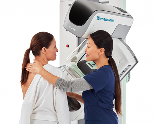
How is 3D Mammography different?
3D mammography is an advanced form of breast imaging where multiple images of the breast from different angles are captured and reconstructed into a three-dimensional image set. The 3D mammogram machine creates both 3D images and standard 2D mammogram images because both types of images have some advantages in seeing certain breast abnormalities.
3D mammograms can:
-
-
- Reduce the need for follow-up imaging
- Improve breast cancer detection, especially in dense breast tissue
- Reduce the number of unnecessary biopsies
-